
Makeup
Hemangioma: Symptoms, Diagnosis, and Treatment
Jul
A hemangioma is basically a “too enthusiastic” cluster of blood vessels that forms a lump or mark. Most are benign
(not cancer), and manyespecially in babiesshrink and fade over time. Still, some hemangiomas cause real trouble
depending on size, depth, and location (near an eye, in the airway, on the lip, or inside an organ like the liver).
This guide breaks down hemangioma symptoms, how clinicians diagnose them, and what treatments are commonly used
todaywithout panic, without fluff, and with just enough humor to keep your eyebrows from permanently moving into
the “worried” position.
What exactly is a hemangioma?
“Hemangioma” is a broad term for a benign growth made from blood vessels. People often use it to describe
infantile hemangiomas (the classic “strawberry” birthmark that shows up in early infancy), but hemangiomas can
also occur internally (for example, a liver hemangioma found incidentally on imaging in adults).
Common types you may hear about
-
Infantile hemangioma (IH): The most common vascular tumor of infancy. It often appears in the first
weeks of life, grows quickly for a period, then slowly involutes (shrinks and fades). -
Congenital hemangioma: Present at birth and behaves differently than infantile hemangiomas.
(These are less common and can have distinct growth patterns.) - Deep (subcutaneous) hemangioma: Sits under the skin and can look bluish or like a soft lump.
- Mixed hemangioma: Has both superficial (red) and deep (bluish) components.
-
Liver (hepatic) hemangioma: A benign vascular lesion in the liveroften symptom-free and found by
accident on ultrasound, CT, or MRI.
A key point: not every vascular-looking spot is a hemangioma. Vascular malformations (like venous malformations)
can look similar but behave differently, so an accurate diagnosis matters.
Hemangioma symptoms: what it can look and feel like
Symptoms on the skin
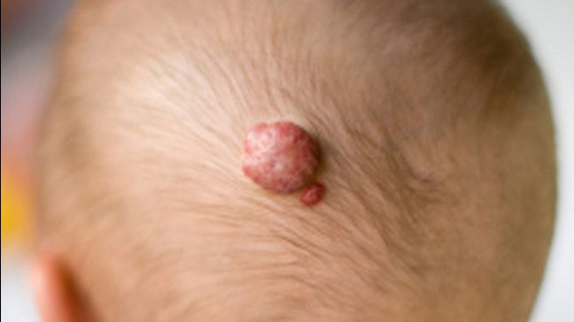
Skin hemangiomas usually show up on the face, scalp, chest, or back, but they can appear anywhere. Superficial
hemangiomas often start as a faint red area and become brighter and more raised. Deep hemangiomas can appear as a
bluish swelling under the skin.
Infantile hemangiomas typically follow a “grow, then glow-down” timeline:
- Early phase: Subtle spot or patch (sometimes mistaken for a scratch or irritation).
- Proliferative (growth) phase: Rapid growth over weeks to months (fastest early in infancy).
- Involution phase: Gradual shrinking/fading over years; the color dulls, the bump softens.
Most are painless, but symptoms can pop up when a hemangioma is in a high-friction area or growing in a sensitive
place. Common complications include:
- Ulceration: The surface breaks down into a soreoften painful and at risk for infection.
- Bleeding: Usually minor, but any persistent bleeding needs medical attention.
- Functional problems: Vision issues (if near the eye), feeding difficulties (near lips/mouth), or
breathing problems (airway involvement). - Cosmetic impact: Larger facial lesions can leave lasting skin changes even after shrinking.
Symptoms when a hemangioma is inside the body
Internal hemangiomas are often discovered incidentallymeaning they show up on imaging done for some other reason.
A classic example is a liver hemangioma found during an ultrasound or MRI. Most cause no symptoms and require no
treatment.
When internal hemangiomas do cause symptoms, it’s usually because they’re large or affecting nearby structures.
For liver hemangiomas, symptoms (when they happen) can include upper abdominal discomfort, feeling full quickly,
bloating, or nausea.
“Don’t wait it out” warning signs
Hemangiomas can be low-drama, but certain situations deserve prompt evaluationespecially in infants. Contact a
clinician soon if you notice:
- A hemangioma near the eye (risk of vision problems).
- Noisy breathing, chronic cough, or breathing difficulty (possible airway involvement).
- Rapid growth, especially on the face or in the diaper area.
- Open sores, crusting, or significant pain (possible ulceration).
- Five or more skin hemangiomas (sometimes linked with liver involvement in infants).
- A large “segmental” hemangioma on the face/scalp (may need evaluation for associated syndromes).
Diagnosis: how clinicians confirm hemangioma
1) History and physical exam (often enough)
Many hemangiomasespecially typical infantile hemangiomascan be diagnosed by appearance and growth pattern alone.
Clinicians consider when it appeared, how quickly it’s changing, whether it’s superficial or deep, and whether
there are any complications (like ulceration or functional impairment).
2) Imaging (used strategically, not automatically)
Imaging is most helpful when the diagnosis isn’t clear, when a lesion is deep, or when clinicians suspect
associated anatomic issues. Common imaging tools include:
-
Ultrasound (often with Doppler): Frequently the first step if the diagnosis is uncertain, and a
common way to evaluate suspected liver hemangiomas. -
MRI: Useful for mapping extent and deeper involvement, or when associated structural
abnormalities are suspected. -
CT: Sometimes used for internal lesions like liver hemangiomas, though MRI is often preferred
for detailed characterization.
3) Extra evaluation for “high-risk” patterns
Some hemangioma patterns are considered high risk because they’re more likely to cause complications or be linked
with other findings. For example:
-
Large segmental facial/scalp hemangiomas: May trigger evaluation for syndromes such as PHACE,
often involving MRI/MRA and other screening. -
Five or more cutaneous hemangiomas in an infant: May prompt screening for liver hemangiomas and,
in certain cases, lab monitoring (including thyroid function if extensive hepatic involvement is suspected).
Translation: clinicians don’t order extra tests to be dramaticthey do it to catch the uncommon-but-important
scenarios early, when treatment can prevent complications.
Treatment: from “watch and wait” to medications and procedures
Step 1: Observation and follow-up (the right move for many)
Most infantile hemangiomas are small, uncomplicated, and self-resolving. In these cases, the plan may be
observation with regular check-insespecially during the rapid growth phase in early infancy. Parents are often
encouraged to take periodic photos (same lighting, same angle) so growth changes are easier to spot.
Observation does not mean ignoring it. It means: “We expect this to improve on its own, and we’re watching for
signs that it won’t.”
Step 2: Beta-blockers (modern first-line therapy for problem hemangiomas)
The biggest change in hemangioma care over the past couple of decades is the use of beta-blockersmedications that
affect blood vessel behavior.
Oral propranolol
Oral propranolol is widely considered the treatment of choice for infantile hemangiomas that need systemic therapy
(for example, lesions threatening vision, breathing, feeding, or those likely to scar significantly). Treatment is
often continued through infancy (commonly until around 1 year of age or longer, depending on response and risk of
rebound growth).
Because propranolol can affect heart rate, blood pressure, and blood sugar, clinicians typically use screening and
monitoring protocolsespecially at the start or when dosing changes. Commonly discussed potential side effects
include low blood sugar, low blood pressure, and wheezing, which is why medical guidance and follow-up are
essential.
Topical timolol
For select small, thin, superficial hemangiomas, clinicians may consider topical timolol (a beta-blocker applied to
the skin). It’s often used when the lesion is superficial and the goal is to reduce redness and growth without
systemic medication.
Step 3: Other medications (when beta-blockers aren’t an option)
If propranolol can’t be used or isn’t effective, clinicians may consider alternatives such as oral corticosteroids
(less common now than in the pre-beta-blocker era), or other therapies depending on the specific case and
specialist input.
Step 4: Laser therapy
Laser treatment may be used for certain hemangiomasoften to help with ulceration care, to reduce persistent
surface redness, or to improve residual visible blood vessels after the hemangioma has involuted. The role of
laser depends on depth, location, and timing.
Step 5: Surgery
Surgery is not the first choice for most infantile hemangiomas during the growth phase. However, it can be helpful
in specific situations, such as removing residual tissue or correcting leftover contour changes after involution,
or when other treatments aren’t suitable. For internal lesions (like certain symptomatic liver hemangiomas), surgery
is uncommon but may be considered in select cases.
Treatment choices by location: why “where” matters as much as “what”
Hemangiomas near the eye (periocular)
A periocular hemangioma can press on the eyelid or distort the eye’s shape, increasing the risk of amblyopia
(“lazy eye”) or other visual development problems. These lesions often require early evaluation and, if indicated,
prompt treatmentcommonly with oral propranolol under medical supervision.
Airway hemangiomas
Hemangiomas in or near the airway can cause noisy breathing (stridor), cough, or respiratory distress. This is a
high-priority scenario that typically involves specialist care and systemic treatment.
Lip, mouth, diaper area, and skin folds
These areas are high-friction zones. Hemangiomas here ulcerate more easily, can be painful, and may complicate
feeding or hygiene. Treatment may focus on preventing ulceration, managing pain, and addressing growth early if
complications are likely.
Liver hemangiomas
In adults, liver hemangiomas are often benign “incidental findings” that don’t need treatment unless symptomatic
or unusually large. In infants, multiple skin hemangiomas can sometimes be associated with liver hemangiomas;
clinicians may screen with imaging and monitor for rare complications (including thyroid dysfunction in extensive
hepatic disease).
Practical, non-medical tips for day-to-day management
- Track changes: Weekly photos during early infancy can help document growth patterns.
-
Protect fragile skin: If the hemangioma is in a rub-prone area, gentle skin care can reduce
irritation (your clinician can recommend appropriate products). - Watch for sores: Ulceration can start as a scab, crust, or “raw” spotbring this up quickly.
-
Plan questions for visits: Ask about risk by location, expected timeline, and what signs mean
“call us sooner.” -
Don’t DIY medications: Beta-blockers are effective but should be used only with medical
oversight.
Prognosis: what happens long term?
Many infantile hemangiomas significantly improve with time, and some leave little trace. Others may leave residual
changes such as extra skin, a faint network of visible blood vessels, or a soft, fibrofatty bump where the lesion
used to be. Early treatment for high-risk lesions can reduce complications and may improve long-term cosmetic and
functional outcomes.
For adult liver hemangiomas, the long-term outlook is usually excellent. Most remain stable and non-problematic.
Follow-up plans vary depending on size, symptoms, and imaging features.
Frequently asked questions
Is a hemangioma cancer?
In typical cases, no. Hemangiomas are benign. That said, diagnosis matters because other vascular lesions can look
similar and may require different care.
Will a baby hemangioma go away on its own?
Many do fade substantially over time. The main reason to treat is not “because it exists,” but because it’s likely
to cause a problem (function, ulceration, scarring risk, or significant disfigurement).
Can hemangiomas come back after treatment?
Some infantile hemangiomas can show rebound growth after stopping medication, which is one reason clinicians may
taper treatment and continue follow-up rather than stopping abruptly.
Experiences: what the hemangioma journey can feel like (and what people often learn)
If you’ve ever watched a baby hemangioma “level up” in real time, you know the emotional whiplash is real. One
week it’s a faint pink smudge. Two weeks later it’s brighter, bumpier, and suddenly every well-meaning stranger at
the grocery store has a theory (“Have you tried… potatoes?”). Families often describe the early months as the
toughestnot necessarily because the hemangioma is dangerous, but because it changes quickly and unpredictably
during the growth phase.
A common parent experience is the “location panic.” A small hemangioma on the torso may feel manageable, while the
same size on the eyelid or lip can feel like an emergency. That’s not overreactingthat’s instinct plus anatomy.
Clinicians worry about the same things: vision development, airway safety, feeding mechanics, and skin breakdown.
Many caregivers report that simply hearing a clear risk explanation (“Here’s what we watch for, and here’s why”)
lowers anxiety more than any generic reassurance.
For families who go the observation route, the experience often becomes a lesson in patience and documentation.
Taking regular photos can turn “I think it’s bigger?” into “Yes, it’s changing, but it’s still within expected
growthand here’s the proof.” Parents also learn that “doing nothing” is rarely the plan. The plan is monitoring,
knowing red flags, and having a fast lane to care if the hemangioma crosses into high-risk territory.
For families who start oral propranolol, the experience is frequently described as a mix of relief and nervousness.
Relief because the hemangioma may soften, lighten, and stop ballooning. Nervousness because it’s a real medication
with real monitoring. Many caregivers say the process becomes easier once they understand the routine:
follow dosing instructions carefully, keep follow-up appointments, and learn practical safety basics (like not
skipping feeds in young infants, since low blood sugar can be a concern with beta-blockers). What stands out in
many stories is how quickly fear becomes confidence when families feel supported and informed.
Ulceration, when it happens, is often the most stressful chapter. Parents describe it as painful to look at and
heartbreaking because the baby may be uncomfortable. The day-to-day then shifts to wound care, protecting the area
from friction, and staying close to the care team. The upside is that ulceration is a well-recognized complication,
and treatment plans are often very practical: manage the sore, reduce irritation, and address the hemangioma’s
growth drivers (sometimes with medication, sometimes with adjunct procedures depending on the case).
Adults who learn they have a liver hemangioma often have a different emotional curve: the scan report sounds scary,
but the follow-up conversation is usually reassuring. Many describe a “doom-scroll moment” (we’ve all been there),
followed by relief when a clinician explains that most liver hemangiomas are benign, common, and simply watched
especially if they’re not causing symptoms. The most helpful coping strategy people report is asking the right
questions: “What features make you confident it’s a hemangioma? Do I need follow-up imaging? What symptoms should
I report?”
Across ages and types, one theme keeps repeating: hemangioma care is less about a single perfect decision and more
about matching the plan to the risk. Low-risk lesions often do well with watchful waiting. Higher-risk lesions do
best when evaluated early and treated thoughtfully. And in both situations, the best outcomes tend to happen when
families have a clear roadmap, not a stack of confusing internet anecdotes.
Conclusion
Hemangiomas are common, usually benign, and often self-resolvingespecially infantile hemangiomas. But “usually”
isn’t the same as “always,” and location matters. The most important steps are recognizing high-risk signs,
getting an accurate diagnosis, and using the right level of treatmentranging from observation to beta-blocker
therapy (like oral propranolol or topical timolol), and occasionally laser or surgery.
If you’re dealing with a hemangioma (for yourself or a child), the goal isn’t to chase perfectionit’s to protect
function, prevent complications, and support healthy healing over time. And yes, you’re allowed to take a deep
breath. Preferably more than one.

